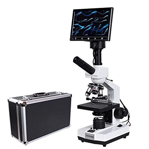


Upon examining the reproductive cells of canines, one may notice several distinctive features. Typically, these cells appear as a whitish or creamy fluid, often with a viscous texture. The consistency can vary, influenced by factors such as the dog’s health, age, and breed.
Microscopically, these cells exhibit a unique structure. Each cell is characterised by a head, midpiece, and tail, which collectively facilitate motility. The head contains genetic material, while the midpiece provides energy for movement, and the tail propels the cell forward. Observing these cells under a microscope reveals an active and dynamic environment, where many of them are constantly swimming and competing for the opportunity to fertilise an egg.
Collecting a sample for examination should be approached with care and professionalism. If you’re considering this for breeding purposes, consulting a veterinarian is advisable. They can provide guidance on proper collection methods and ensure the health and viability of the sample. Understanding these aspects not only aids in responsible breeding practices but also enhances one’s knowledge about canine reproduction.
Visual Characteristics of Canine Reproductive Cells Under Microscope
To properly assess the reproductive cells of canines, a microscope is essential. These cells display distinctive features that are critical for understanding fertility. Under magnification, you will observe the following characteristics:
| Characteristic | Description |
|---|---|
| Shape | Typically, these cells exhibit an elongated structure, resembling a tadpole. The head is oval, while the tail is long and whip-like, aiding in movement. |
| Size | These reproductive cells are relatively small, measuring approximately 60-70 micrometres in length, with the head being about 5-6 micrometres wide. |
| Motility | Healthy specimens demonstrate vigorous swimming patterns. Rapid and straight movements indicate good viability, while sluggish or erratic motion suggests potential issues. |
| Colour | Under the microscope, these cells usually appear translucent or slightly opaque, which can vary based on the sample’s health and concentration. |
| Aggregation | These cells may sometimes cluster together, which could indicate potential fertility challenges or sample handling issues. |
Regular evaluation of these characteristics is beneficial for breeders and pet owners alike. Understanding the physical traits of reproductive cells can aid in making informed decisions regarding mating and overall health. For further insights on canine training and welfare, consider exploring whether is training a dog with a shock collar bad.
Comparing Canine Reproductive Cells to Other Species
In terms of morphology, the reproductive cells of canines exhibit distinct features, setting them apart from those of various animals. For instance, the head of a canine reproductive cell is oval-shaped, which differs from the elongated head seen in equine cells. This shape influences motility patterns, with canine cells demonstrating a unique swimming style that can be observed under a microscope. Their tail structure, long and whip-like, contributes to their vigorous movement in the reproductive tract.
Size Variations Across Species
When examining dimensions, canine reproductive cells typically measure around 60 micrometres in length. In contrast, feline cells are slightly smaller, averaging around 50 micrometres. This size difference may play a role in their respective fertilisation rates and reproductive strategies. Larger cells, as seen in some livestock breeds, can exceed 70 micrometres, which may enhance their chances of reaching the egg in larger reproductive tracts.
Behavioural Characteristics
The motility rate of canine reproductive cells generally hovers around 70 to 80%, which is considered quite robust. In comparison, bovine cells exhibit lower motility, often ranging between 40 to 60%. This disparity can affect fertilisation success rates, with more motile cells having a better chance of successful fertilisation. Environmental factors, such as temperature and pH levels, also influence the activity of these cells, making understanding their behaviour crucial for successful breeding practices.
Factors Affecting the Appearance of Canine Reproductive Cells
The appearance of canine reproductive cells can vary significantly based on several key factors. One major influence is the age of the animal. Young males typically exhibit healthier and more motile cells compared to older ones, whose reproductive health may decline over time.
Nutrition plays a vital role as well. A balanced diet rich in essential fatty acids, zinc, and antioxidants contributes positively to reproductive health. Dogs fed high-quality commercial diets tend to show improved cell quality, while those on poor diets may produce suboptimal samples.
Environmental conditions cannot be overlooked. Exposure to toxins, pollutants, or extreme temperatures can adversely affect reproductive health. Keeping the living environment clean and safe is crucial for maintaining optimal conditions for male reproductive function.
Stress levels also impact the quality of reproductive cells. High-stress situations, such as loud noises or changes in routine, can lead to decreased motility and viability. Ensuring a calm and stable environment helps mitigate these effects.
Health issues, such as infections or hormonal imbalances, can significantly alter the appearance of reproductive cells. Regular veterinary check-ups and prompt treatment of any health concerns are essential for maintaining reproductive viability.
Finally, genetic factors can influence the characteristics of reproductive cells. Certain breeds may have inherent traits that affect the appearance and quality. Understanding breed-specific traits can provide insights into what to expect and how to improve overall health.
Common Misconceptions About Canine Reproductive Cells’ Appearance
Many people envision reproductive cells as large or distinctly visible entities, but that’s far from reality. These cells are microscopic and require specialised equipment to observe. A common myth is that they have a uniform shape. In truth, individual variations exist, influenced by health and genetics.
Another misconception is that these cells are all motile. While many are indeed active, a significant percentage may be non-motile or exhibit reduced motility. This variability can lead to misunderstandings about fertility and breeding potential. Some believe that a high concentration of these cells guarantees successful mating, but quality often trumps quantity. Healthy, active cells are far more crucial than sheer numbers.
Many assume that colour plays a role in assessing health. However, the appearance under a microscope is typically a translucent or slightly opaque hue, which does not inherently indicate quality. Misinterpretations of colour can lead to unnecessary concerns about reproductive health.
Additionally, there’s a belief that these cells appear similar across all breeds. In reality, variations can be observed based on breed characteristics. This can affect motility and morphology, making it essential for breeders to understand their specific breed’s reproductive traits.
Lastly, there’s an idea that stress does not impact these cells. In fact, environmental factors, stress, and overall health significantly influence the morphology and motility of these reproductive cells. Keeping breeding animals in a calm, healthy environment can enhance reproductive success.
Collecting and Examining Canine Semen Safely
Use a sterile collection device, such as a plastic artificial vagina or a clean glass container. Ensure your canine is comfortable; a relaxed environment can make the process smoother. If possible, have a familiar partner present to stimulate interest. This reduces stress and encourages natural behaviour.
Preparation for Collection
Ensure all equipment is sterile. Before collection, wash your hands thoroughly and wear disposable gloves to maintain hygiene. If you’re using an artificial vagina, warm it to body temperature. This mimics the natural conditions and can encourage successful collection.
Examination Techniques
After collection, examine the fluid quickly to assess quality. Use a microscope for a detailed analysis. Place a small drop on a slide, cover it gently with a cover slip, and observe under low magnification. Look for motility and morphology, as these factors are crucial for assessing reproductive health.
Importance of Sperm Quality in Dog Breeding
Prioritising high-quality reproductive material is paramount for successful breeding. The genetic health and overall vitality of the offspring depend significantly on the characteristics of the male’s reproductive cells. A few key aspects should be considered to ensure optimal results.
Key Factors in Quality Assessment
- Motility: Active and vigorous movement is a clear indicator of healthy reproductive cells. Ideally, over 70% of the cells should exhibit strong motility.
- Morphology: The shape and structure of the cells play a crucial role in fertility. Abnormal shapes can lead to fertilisation issues.
- Concentration: A higher concentration of reproductive cells increases the likelihood of successful mating. A minimum count of 300 million per millilitre is often recommended.
Implications of Quality on Breeding Outcomes
High-quality reproductive cells lead to healthier litters with improved genetic traits. Inferior quality can result in lower conception rates, increased risk of genetic disorders, and fewer viable offspring. Breeders should conduct thorough evaluations before selecting a male for breeding. Regular health checks and semen analysis can reveal underlying issues that may affect fertility.
Utilising advanced techniques for collection and analysis enhances the accuracy of assessments. Collaborating with a veterinarian specialising in reproductive health can provide invaluable insights and guidance. By focusing on these factors, breeders can significantly enhance the success of their breeding programmes and contribute positively to the breed’s future.
Signs of Abnormalities in Canine Reproductive Cells
Identifying irregularities in reproductive cells is crucial for assessing male fertility. Here are key indicators to watch for:
Visual Indicators
- Motility Issues: Healthy samples should show vigorous movement. Reduced activity or sluggishness can indicate problems.
- Abnormal Morphology: Look for unusual shapes. Common abnormalities include double tails, misshapen heads, or flattened bodies.
- Clumping: Healthy specimens usually appear individually. Clumping may signal an underlying issue.
- Colour Changes: A normal sample is typically milky or creamy. Yellow or brown hues might suggest infection or contamination.
Laboratory Analysis
In addition to visual checks, laboratory tests can provide deeper insights:
- Hormonal Imbalances: Blood tests can reveal hormonal levels that affect fertility.
- DNA Fragmentation: High levels of DNA damage can impact successful fertilisation.
- Concentration Levels: Low concentration may indicate a production issue.
Regular evaluations can help detect problems early, which is essential for successful breeding. If abnormalities are suspected, consulting a veterinary specialist is advisable for thorough testing and guidance.


